

Author Ken Walker ◦
I’m glad you enjoyed the 80 stack single image from Gary Taylor’s bee.
My research work occurs mainly in the world of under the microscope and I am privileged to see animals (in this case bees) in ways that most people could not imagine. I see them “warts and all” so I thought I would take a few more 80 image stack photos and show you what the head of Gary’s bee looks like at high magnification and then a general dorsal and lateral view for overall views.
Hope you enjoy these images. I have marked the original head image stack with arrows and numbers of where I took high detailed images from the head of Gary’s specimen. The additional photos will appear in separate posts so I can explain what you are seeing.
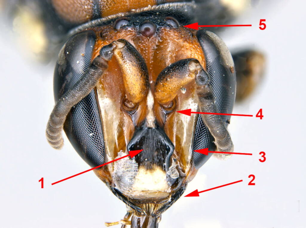
Image 1. this is a view of the clypeus and supraclypeal area of the bee. Basically this is the bee’s top lip. Notice the vertical striations. Usually striations are designed to increase the surface area compared to a smooth surface so the male bee obviously uses his top lip for some function. Perhaps to push other males away from the female so he can mate.
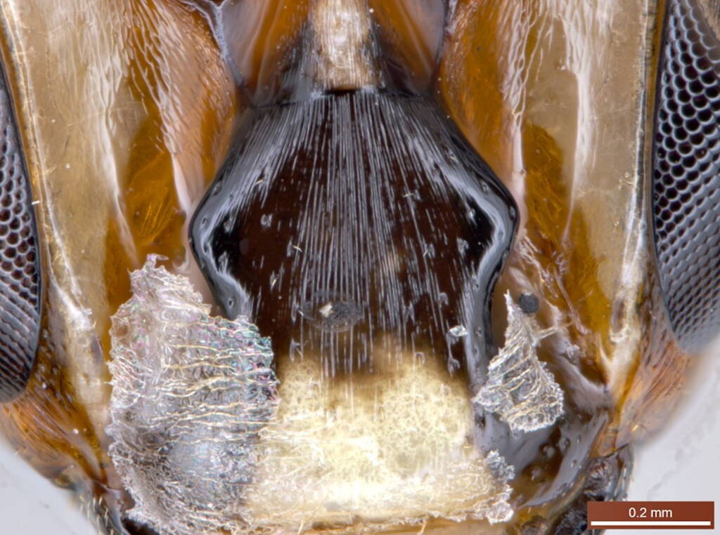
Image 2. This is the left mandible or jaw of the bee. Notice the ridges that run the length of the mandible. Also note the gap between the top of the mandible and the base of the compound eye. This area is called the “malar space”. In this case, the space is quite large whereas in other bees there is no space. Also notice the setae running across the anterior margin of the clypeus – a bit like a moustache.
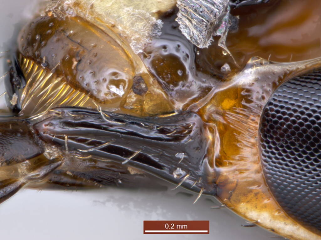
Image 3. This image shows the line between the edge of the compound eye and the inner margin of the face that meets the eye. This line is always just perfect.
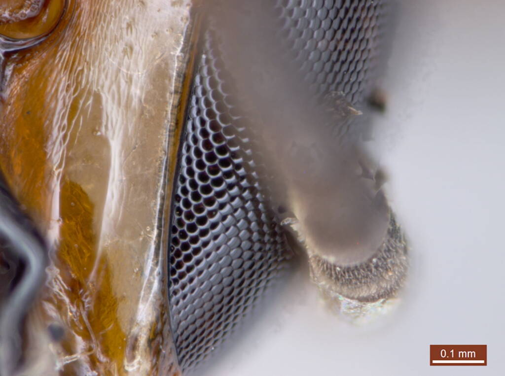
Image 4. This image shows the bases of the two antennae. Notice how the base is rounded and inserted into a socket so the antennae can swivel and move around in most directions. The space between the antennal bases is an important character to distinguish between different species.
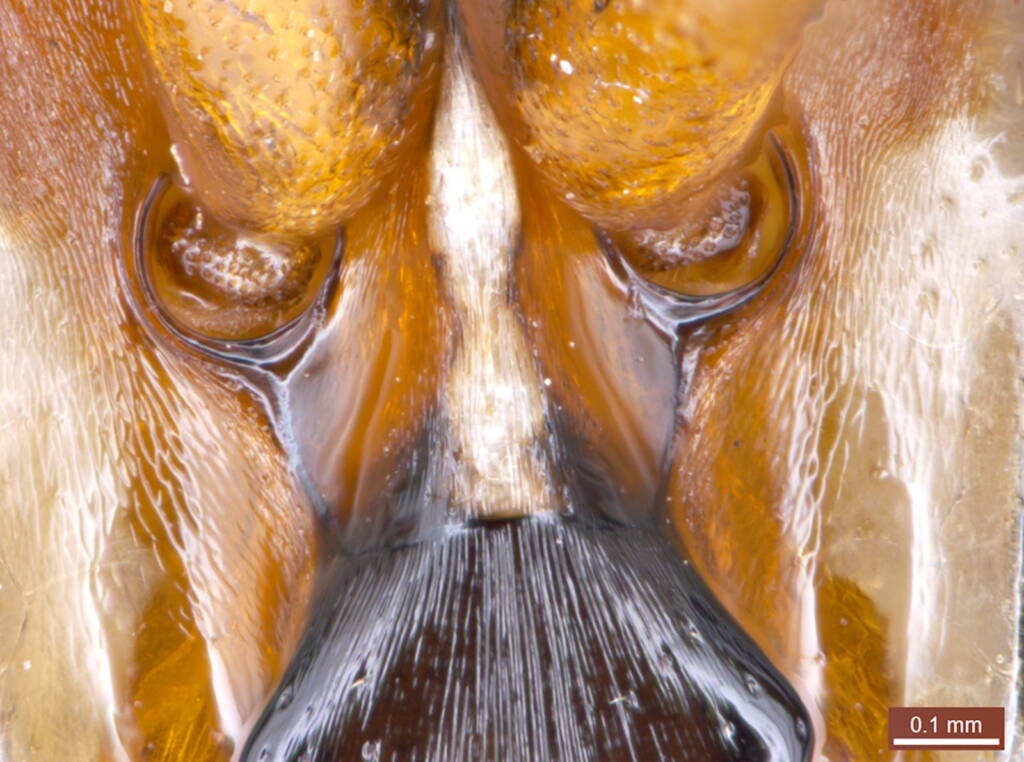
Image 5. Finally, this image was taken at the top or vertex of the head and shows the 3 ocelli or additional eyes arranged in the shape of a triangle.
Note that while the 2 large eyes on each side of the head are compound or made up of many small eyes (called ommatidia), each ocellus is a single reflex lens. Ocelli are eyes but with different functions to the 2 large eyes. Ocelli help bees with two main functions. One is orientation. When the bee flies away from the nest, it needs to be able to find its way back. The ocelli triangulates with the sun and time of day and are the bee’s navigation guide on how to retrace his or her steps. Almost all insects have ocelli. The second value of the ocelli is that they tell the bee what season of the year it is. If the daylengths are increasing then the season is going towards summer and bees are out making and provisioning nests. If the daylengths are decreasing, then the bees know that winter is approaching and they need to complete provisioning and sealing nests as when winter comes all of the adult bees will die.
So, insects generally have 5 eyes – 2 large compound eyes on each side of the head used for seeing and 3 small single lens eyes called ocelli that allow be the bee to navigate and estimate the time of season. Very cool.
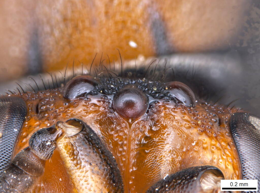
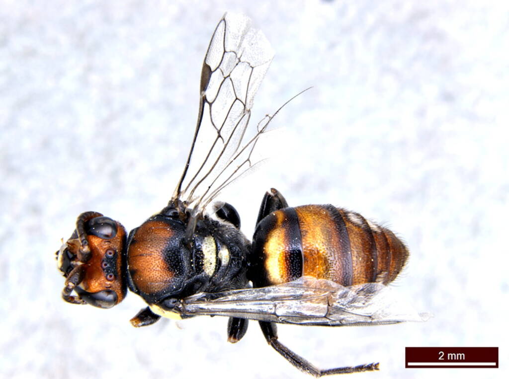
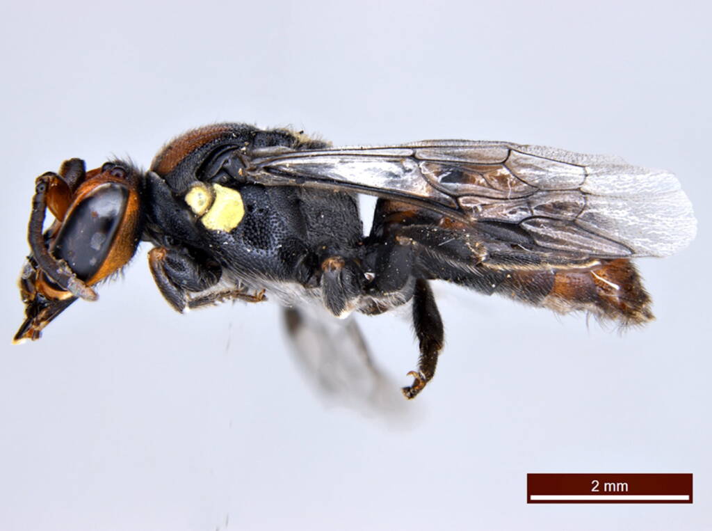
Image 6. And finally, the dorsal and lateral views of Gary’s bee.
Bye.
I noticed some debris hanging out of one of the nests | “warts and all”
Footnote & References
- All photographs of the male Meroglossa rubricata © Ken Walker
- Male Meroglossa rubricata supplied specimen by Gary Taylor